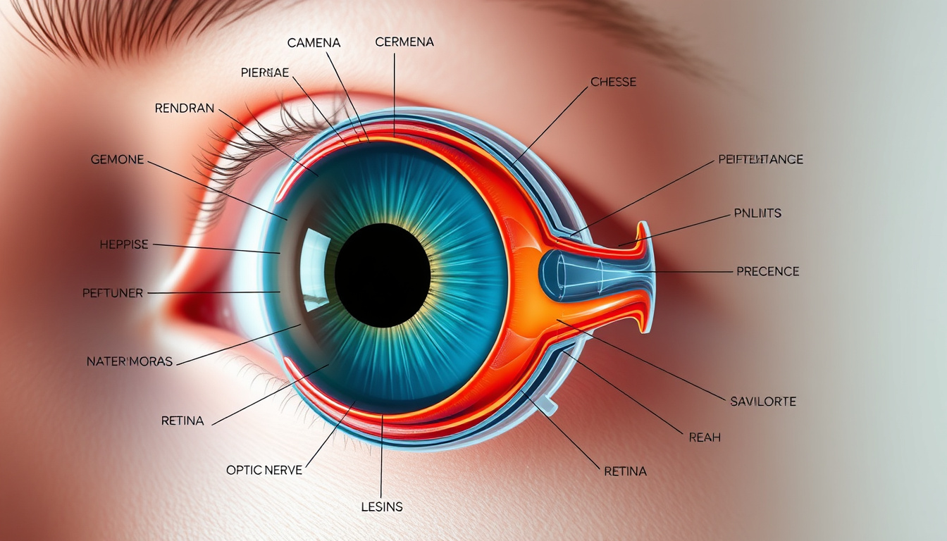
You probably don’t think about your eyes until something goes wrong, but every blink, glance, and squint depends on a finely tuned team of parts working together. The parts of the eye are less like simple components and more like a precision instrument: the cornea bends incoming light, the iris adjusts exposure, and the lens fine-tunes focus before the retina converts everything into signals your brain can read.
Main parts commonly described: 10+ ·
Average adult eye diameter: 24 mm ·
Number of eye layers: 3 (sclera, choroid, retina) ·
Corneal thickness: 0.5 mm
Quick snapshot
- Clear dome protects the eye (Merck Manuals clinical reference)
- Bends light to focus (Merck Manuals clinical reference)
- No blood vessels; gets oxygen from air (Merck Manuals clinical reference)
- Iris is the colored part (AboutKidsHealth pediatric reference)
- Pupil is the opening (AboutKidsHealth pediatric reference)
- Iris controls light entry (AboutKidsHealth pediatric reference)
- Transparent and flexible (Dr. Sabrina clinical blog)
- Changes shape to focus (Dr. Sabrina clinical blog)
- Clouding leads to cataracts (Dr. Sabrina clinical blog)
- Light-sensitive layer (AboutKidsHealth pediatric reference)
- Rods for dim light, cones for color (AboutKidsHealth pediatric reference)
- Converts light to nerve signals (AboutKidsHealth pediatric reference)
Five key structures, one pattern: each part of the eye has a single, irreplaceable job, and the chain breaks if any link fails.
“The sclera runs continuously from the cornea at the front to the optic nerve at the back, providing structural support.” — Cleveland Clinic medical reference
| Structure | Key Fact |
|---|---|
| Toughest outer layer | Sclera (Cleveland Clinic medical reference) |
| Main focusing element | Cornea (provides 2/3 of focusing power) |
| Adjustable focusing structure | Lens (Dr. Sabrina clinical blog) |
| Light-sensitive tissue | Retina (AboutKidsHealth pediatric reference) |
| Nerve connection to brain | Optic nerve |
What Is the White Part of the Eye Called?
The white part of your eye has a real name: the sclera. It forms the tough, fibrous outer shell that gives the eyeball its round shape and protects the delicate inner structures. According to the Cleveland Clinic medical reference, the sclera runs continuously from the cornea at the front to the optic nerve at the back, providing structural support.
The sclera and its protective function
- The sclera is composed of dense collagen and elastic fibers, making it rigid enough to withstand minor impacts.
- A clear, thin layer called the conjunctiva covers the visible part of the sclera and keeps it moist.
- Four of the six extraocular muscles attach directly to the sclera, enabling eye movement.
Common conditions affecting the sclera
- Episcleritis: inflammation of the tissue just above the sclera, often causing redness and discomfort.
- Scleritis: deeper, more painful inflammation that can be linked to autoimmune diseases.
- Jaundice: a yellow tint to the sclera signals liver problems and requires medical attention.
The takeaway: the sclera is your eye’s structural scaffold and its first line of defense—when it changes color or becomes painful, something deeper is likely going on.
What Are the Main Parts of the Eye and Their Functions?
Six main parts form the core of how we see. Each one plays a distinct role, and they work in sequence the moment light enters the eye.
Cornea: the clear front window
- The cornea provides approximately two-thirds of the eye’s total focusing power.
- It is avascular—meaning it has no blood vessels—and receives oxygen directly from the air.
- According to the Merck Manuals clinical reference, the cornea refracts light as it enters, making it the eye’s primary optical component.
Iris and pupil: controlling light
- The iris is the pigmented part that gives your eye its color—brown, blue, green, or hazel.
- At its center lies the pupil, a black circular opening that widens in the dark and narrows in bright light.
- The AboutKidsHealth pediatric reference explains that the iris adjusts pupil size to regulate light intake, much like a camera aperture.
Lens: focusing light
- Behind the pupil sits the lens, a flexible, transparent structure that changes shape to fine-tune focus.
- When you look at something close, the lens thickens; for distant objects, it flattens.
- As Dr. Sabrina’s clinical blog notes, the lens’s elasticity gradually decreases with age—this is why many people eventually need reading glasses.
Retina: converting light to signals
- The retina lines the back of the eye like film in a camera, containing millions of photoreceptor cells.
- Rods handle low-light and peripheral vision; cones manage color and fine detail in bright light.
- According to the AboutKidsHealth pediatric reference, the retina converts light into electrical impulses and sends them along the optic nerve.
Optic nerve: transmitting to brain
- The optic nerve acts as the data cable, carrying visual signals from the retina to the brain’s visual cortex.
- Damage to the optic nerve—from glaucoma, trauma, or disease—can cause irreversible vision loss.
- Dr. Sabrina’s clinical blog emphasizes that once optic nerve fibers die, they do not regenerate.
What this means: each part of the eye is optimized for one purpose, and the system’s elegance lies in that precise division of labor.
“The cornea is the eye’s primary optical component, acting as a clear window that protects the eye and bends incoming light to focus it.” — Merck Manuals clinical reference
What Are the Parts of the Outer Eye?
The outer eye includes structures that protect, lubricate, and move the eyeball—often overlooked until irritation sets in.
Eyelids and lashes: protection
- Eyelids act as rapid shutters, distributing tears across the surface and sweeping away debris with every blink.
- Eyelashes form a physical barrier that triggers a reflexive blink when touched by dust or insects.
- The News-Medical.Net anatomy reference describes eyelids as primary barriers against the external environment.
Conjunctiva: lubrication
- This clear, thin membrane covers the visible sclera and lines the inside of the eyelids.
- It contains tiny glands that produce mucus and tears to keep the eye moist and free of infection.
- When the conjunctiva becomes inflamed—pink eye or conjunctivitis—it causes redness, itching, and discharge.
Extraocular muscles: movement
- Six muscles control each eye, allowing smooth tracking, rapid shifts, and coordinated binocular vision.
- These muscles are among the fastest-contracting in the human body, moving the eye in milliseconds.
- Imbalance in these muscles leads to strabismus (crossed eyes) or double vision.
The trade-off: protective structures sacrifice some of the visual field for safety, but that’s a bargain worth making.
How Do the Parts of the Eye Work Together to Allow Vision?
Vision is a sequence, not a snapshot. Light travels through five distinct zones before your brain interprets the image.
The visual pathway: light to brain
- Light enters through the cornea, which bends (refracts) the rays toward the pupil.
- The iris adjusts the size of the pupil to control the amount of light entering.
- The lens fine-tunes focus, changing shape to bring the image to a sharp point on the retina.
- The retina’s photoreceptors convert the focused light into electrical signals.
- The optic nerve transmits those signals to the visual cortex at the back of the brain.
Role of the vitreous humor
- The vitreous humor is a clear, jelly-like substance that fills the large chamber behind the lens.
- According to the Cleveland Clinic medical reference, it maintains the eye’s spherical shape and allows light to pass through without obstruction.
- As people age, the vitreous can shrink and detach, sometimes causing floaters or flashes.
Role of the choroid
- The choroid sits between the retina and sclera, packed with blood vessels that supply oxygen and nutrients to the outer retina.
- It also contains melanin, which absorbs stray light to prevent internal reflections that would blur vision.
- The News-Medical.Net anatomy reference calls it the vascular middle layer that nourishes the retina.
The pattern: light is bent, regulated, focused, converted, and transmitted—in that order, every time you open your eyes.
The average adult eye measures just 24 mm in diameter, yet this small organ contains more than 120 million rods and 6 million cones at the retina. When any part of this chain fails—a cloudy lens, a damaged optic nerve, a detached retina—the entire visual system breaks down.
What Is a Simple Eye Diagram for Kids?
Simplifying the eye for young learners means stripping away medical jargon and focusing on what each part does in everyday language.
Labelling the basic parts
- A child-friendly diagram includes the cornea, pupil, iris, lens, retina, and optic nerve as the six main labels.
- Each part can be drawn in sequence: light enters the front (cornea), passes through the colored ring (iris), goes through the dark hole (pupil), gets bent by the clear crystal (lens), and lands on the screen (retina).
- The optic nerve is drawn as a wire connecting the eye to the brain, like a charging cable.
Simplified functions for Grade 3
- Cornea: “The clear window that lets light in.”
- Iris: “The color ring that controls how much light enters.”
- Pupil: “The black hole that gets bigger in the dark.”
- Lens: “The part that bends to focus, like a camera zoom.”
- Retina: “The screen where the picture forms.”
- Optic nerve: “The cable that sends the picture to your brain.”
For educators, the key is to use analogies kids already understand: the lens works like a camera zoom, the iris works like a door, and the optic nerve works like a USB cable from the eye to the brain.
Related reading: Sunglasses Store Near Me: Top Dublin Shops & Prices · Flu Vaccine Side Effects: Normal vs Serious Reactions
Frequently asked questions
Is the pupil a hole or a structure?
The pupil is not a physical structure—it is the open space at the center of the iris. The iris expands and contracts around this opening to control light entry. According to the News-Medical.Net anatomy reference, the pupil itself is simply the black circular opening that widens in dark environments.
What part of the eye gives color?
The iris is the pigmented part of the eye that determines its color—brown, blue, green, or hazel. The amount and type of melanin in the iris tissue dictate the shade. As the AboutKidsHealth pediatric reference explains, the iris is the colored ring that surrounds the pupil.
Can the lens be replaced?
Yes. Cataract surgery involves removing the clouded natural lens and replacing it with a clear artificial intraocular lens (IOL). It is one of the most common and successful surgical procedures worldwide, restoring vision in millions of patients each year.
What is the macula and what does it do?
The macula is the small central area of the retina responsible for sharp, detailed central vision. It enables reading, driving, and recognizing faces. As Dr. Sabrina’s clinical blog notes, damage to the macula—as in age-related macular degeneration—affects central vision while leaving peripheral sight intact.
Why is the back of the eye dark?
The back of the eye appears dark because the choroid layer is rich in melanin, a pigment that absorbs stray light. This prevents internal light scattering that would reduce the clarity of the visual image projected onto the retina.
What are the three chambers of the eye?
The eye has three chambers: the anterior chamber (between the cornea and iris), the posterior chamber (between the iris and lens), and the vitreous chamber (between the lens and retina). The anterior and posterior chambers are filled with aqueous humor, while the vitreous chamber contains the gel-like vitreous humor.
How many extraocular muscles are there?
There are six extraocular muscles attached to each eye: four rectus muscles (superior, inferior, medial, lateral) and two oblique muscles (superior and inferior). These muscles work together to move the eye in every direction, allowing smooth tracking and rapid shifts of gaze.
What fluid fills the front of the eye?
The anterior part of the eye is filled with aqueous humor, a clear, watery fluid that provides nutrients to the cornea and lens and maintains intraocular pressure. According to the Cleveland Clinic medical reference, aqueous humor is produced in the ciliary body and drains through the trabecular meshwork.
For anyone curious about their own vision, the takeaway is simple: the cornea handles the first big bend of light, the lens adjusts the fine focus, and the retina translates everything into signals your brain can read. Problems arise when any one of these parts of the eye stops doing its job—whether through injury, aging, or disease. For parents teaching kids about the eye, the camera analogy works: cornea as the lens cap, iris as the aperture, lens as the zoom, and retina as the digital sensor. For students or professionals who need a deeper grasp, the clinical reality is that each structure is irreplaceable. The implication for anyone who relies on their sight (which is all of us): learn the parts of the eye, understand what each one does, and you will immediately recognize when something feels off—and know exactly which specialist to see.